
2D Echocardiography (also known as 2D Echo) is a non-invasive imaging technique used to visualize the heart's structure and function. It uses ultrasound waves to create two-dimensional images of the heart, allowing physicians to assess various cardiac functions, including the size, shape, and movement of the heart's chambers, valves, and walls. This technique is commonly used to diagnose and monitor a variety of heart conditions.
Key features of 2D Echo Cardiography :
2D echocardiography (2D echo) is a type of ultrasound imaging used to visualize the heart in real time. It provides valuable diagnostic information about the structure and function of the heart. 2D echocardiography is a non-invasive, widely used diagnostic tool that provides critical insights into heart health and guides treatment decisions in cardiology.
Assessment of Heart Structure :
2D echo can evaluate the size, shape, and thickness of the heart chambers (atria and ventricles).
It helps in assessing the structure and movement of the heart valves (mitral, aortic, tricuspid, and pulmonary) to detect conditions like stenosis or regurgitation (leaking).
It can visualize the pericardium, the sac surrounding the heart, to detect conditions like pericardial effusion (fluid accumulation).
Evaluation of Heart Function :
Ejection Fraction (EF) : 2D echo is commonly used to measure the ejection fraction, which is the percentage of blood pumped out of the left ventricle with each heartbeat. It helps in diagnosing heart failure.
Wall Motion Abnormalities : It helps detect abnormalities in the motion of the heart walls, which may indicate previous heart attacks or ischemia (lack of blood flow).
Detection of Heart Diseases :
Congenital Heart Defects : 2D echo is used to identify structural abnormalities present at birth, such as atrial or ventricular septal defects.
Cardiomyopathy : It can detect conditions like dilated or hypertrophic cardiomyopathy (enlarged or thickened heart muscles).
Infective Endocarditis : It helps in identifying vegetations or infections on heart valves.
Assessing Heart's Response to Treatments :
It is often used in monitoring the heart’s response to treatments like medication, surgical procedures, or device implantation (e.g., pacemakers or defibrillators).
Guiding Interventions :
Guiding Cardiac Procedures : 2D echo is used during certain procedures like catheter-based interventions, valve replacements, and during the implantation of pacemakers.
Screening for Pulmonary Hypertension :
By assessing the right ventricle and estimating pulmonary artery pressures, 2D echo helps in detecting pulmonary hypertension (high blood pressure in the lungs).
Procedure of 2D Echo cardiography :
2D Echocardiography : is a non-invasive imaging technique used to visualize the heart's structures and assess its function. This procedure is safe, painless, and commonly used to diagnose and monitor heart conditions like heart failure, valve disorders, and congenital heart diseases.
Patient Preparation : The patient is usually asked to remove clothing from the upper body and change into a hospital gown. The patient lies on an examination table, typically on their left side, to bring the heart closer to the chest wall. Sometimes, small electrode patches may be placed on the patient’s chest to monitor the heart’s electrical activity (ECG).
Application of Gel : A water-based gel is applied to the chest. This helps in conducting sound waves by removing any air between the skin and the transducer.
Transducer Placement : The transducer (a small handheld device) is placed on the chest over the heart area. The transducer emits high-frequency sound waves (ultrasound) that penetrate the body.
Image Acquisition : The technician or cardiologist moves the transducer around the chest area to obtain different views of the heart.
Parasternal View provides a view of the heart's chambers and valves. Apical View shows the heart from the bottom, providing a good view of the left ventricle and mitral valve. Subcostal View shows the heart from beneath the rib cage. Suprasternal View is used to visualize large vessels, such as the aorta.
Image Interpretation : The machine converts the sound waves into real-time moving images of the heart. The cardiologist evaluates: Heart chambers’ size and function. Heart valve structure and function. The movement of the heart muscle. Presence of fluid around the heart (pericardial effusion). Structural abnormalities like septal defects or masses.
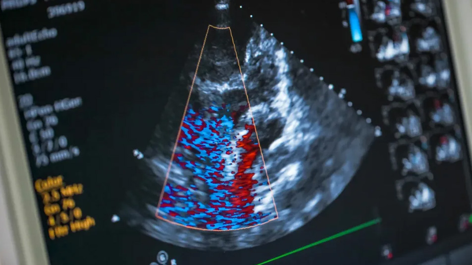
Color Doppler (if used) : Color Doppler imaging can be added to assess blood flow across the heart’s valves and chambers. It helps identify abnormal flow patterns such as regurgitation or stenosis.
Duration : The procedure typically takes 15 to 30 minutes, but it might take longer if more detailed views are needed.
Post-procedure : The gel is wiped off the patient’s chest, and they can resume normal activities immediately. The cardiologist reviews the images and prepares a report for diagnosis or further treatment.
Precautions before Echo Cardiography :
Before undergoing an echocardiography (echo), it is important to follow certain precautions to ensure the test results are accurate and the procedure goes smoothly. Here are some things to avoid :
Avoid Eating or Drinking (in certain cases) :
Fasting : If you are having a transesophageal echocardiogram (TEE), you may be asked to fast for 6-8 hours before the test. This is because the procedure involves inserting a probe down your throat, and fasting minimizes the risk of aspiration (food or liquid entering the lungs).
For a transthoracic echocardiogram (TTE) (the most common type, using an ultrasound probe on your chest), fasting is generally not required.
Avoid Caffeine :
Caffeine can increase your heart rate, which may affect the results. It's generally advised to avoid caffeinated beverages like coffee, tea, soda, or energy drinks for at least 24 hours before the test.
Avoid Smoking or Nicotine :
Smoking can increase heart rate and affect blood flow, which could impact the accuracy of the test. Avoid smoking or using nicotine products for at least a few hours before the test.
Avoid Strenuous Exercise :
Avoid intense physical activity before the procedure, as it can elevate your heart rate and potentially affect the readings during the test.
Medication Considerations :
Some medications can affect heart rate or blood pressure. Follow your doctor’s instructions regarding medications. They may ask you to avoid certain heart medications (e.g., beta-blockers) before the test or, in some cases, continue them as normal.
Avoid Lotions or Creams on Your Chest :
Do not apply any lotions, creams, or oils on your chest before the test, as these can interfere with the ability of the ultrasound probe to make proper contact with your skin.
Following these guidelines will help ensure that your echocardiogram results are accurate and reliable. Always consult your healthcare provider for specific instructions based on your medical condition and the type of echocardiography being performed.